
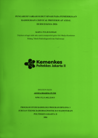
Karya Tulis Ilmiah
PENGARUH VARIASI SUDUT SINAR PADA PEMERIKSAAN RADIOGRAGFI CERVICAL PROYEKSI AP AXIAL DI RSUD KOJA 2026
Anteroposterior (AP) axial cervical spine radiography is one of the radiographic examinations used to visualize the anatomical structures of the cervical vertebrae. In this context, variations in X-ray beam angulation during this examination may affect the quality of the resulting anatomical visualization. Therefore, an evaluation is consequently required to determine the beam angulation that provides an optimal image quality for the visualization of cervical spine anatomy.\r\nThe objective of this research was to describe the procedure for anteroposterior (AP) axial cervical spine radiography performed in the Radiology Department of Koja Regional General Hospital and to analyse the differences in the quality of cervical spine anatomical visualization using 10° and 15° cranial X-ray beam angulations. This research applied a quantitative method with a comparative experimental and observational design. As for the research, it was conducted in Radiology Department of Koja Regional General Hospital from January to April 2026 and included a sample of 13 patients. Radiographic examinations were performed using the anteroposterior (AP) axial projection with central ray angulations of 10° and 15° cranially. Furthermore, the data were entirely collected through a literature review, direct observation, and experimental procedures. The research instruments included a Digital Radiography (DR) system, a Picture Archiving and Communication System (PACS), an observation checklist, and a research data collection form. Data processing consisted of editing, coding, data entry, and data cleaning, followed by statistical analysis performed at a 95% confidence level.\r\nThe results of research showed that the AP axial cervical spine radiographic examination undertaken at Koja Regional General Hospital was performed with the patient in the erect position, with the central ray directed to the fourth cervical vertebra (C4). Based on the observational findings and statistical analysis, a p-value of < 0.05 was obtained, indicating a statistically significant difference in the quality of cervical spine anatomical visualization between the 10° and 15° cranial beam angulations. A 15° cranial beam angulation produced clearer visualization of the vertebral bodies and intervertebral disc spaces than a 10° cranial beam angulation, thereby providing superior overall image quality.\r\nKeywords : Cervical Spine; AP Axial; Cranial Beam Angulation; Vertebral Bodies; Intervertebral Disc Spaces.
Ketersediaan
Informasi Detail
- Judul Seri
-
-
- No. Panggil
-
001.43 ASY p
- Penerbit
- Jakarta : Diploma Tiga Radiologi., 2026
- Deskripsi Fisik
-
xii + 88 hlm. : ilus. ; 21 x 29,7 cm
- Bahasa
-
Indonesia
- ISBN/ISSN
-
-
- Klasifikasi
-
001.43
- Tipe Isi
-
-
- Tipe Media
-
-
- Tipe Pembawa
-
-
- Edisi
-
-
- Subjek
- Info Detail Spesifik
-
-
- Pernyataan Tanggungjawab
-
Asyifa Dharma Putri
Versi lain/terkait
Tidak tersedia versi lain
Lampiran Berkas
Komentar
Anda harus masuk sebelum memberikan komentar
 Karya Umum
  Filsafat
  Agama
Agama  Ilmu-ilmu Sosial
  Bahasa
  Ilmu-ilmu Murni
  Ilmu-ilmu Terapan
  Kesenian, Hiburan, dan Olahraga
  Kesusastraan
  Geografi dan Sejarah